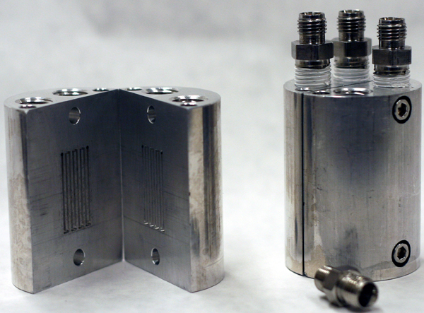
Cylindrical fuel cell used in 3D tomography experiment
The need for non-invasive methods to analyze a polymer electrolyte membrane fuel cell’s (PEMFC) water distribution in both in-situ and ex-situ conditions makes neutron radiography an excellent imaging choice. Neutron imaging is similar but complimentary to x-ray imaging which can penetrate through metal allowing one to observe the membrane and channel features inside a fuel cell. Our group has set out an in-situ neutron tomography system at UC Davis McClellan Nuclear Radiation Center (MNRC), to produce clear 3D tomography PEMFC images and demonstrate further the benefits it has over the 2D techniques.

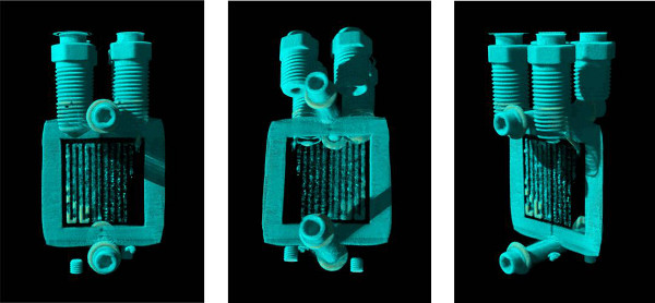
Screen capture of the 3D animation rendered with the reconstructed voxel data set. The internal structure can be analyzed by freely rotating the model. Water blockages inside the flow channel can be immediately identified.
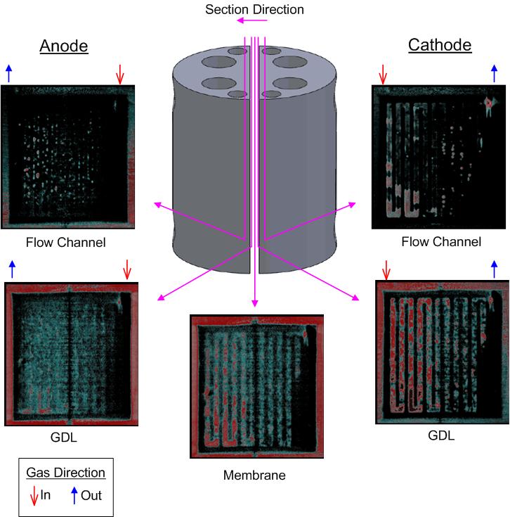
The 3D model created in this work allows for a clear visualization of the fuel cell interior. The model can reconstruct a planar image at an arbitrary plane to provide various cross-sectional views. The following presents liquid water distribution in the flow channels, GDLs and MEA. This capability will provide the fuel cell research community with significant benefit to investigate water and thermal management in a PEM fuel cell.
